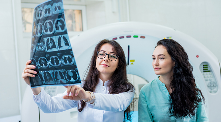
Medical Image Processing
For the common man, Medical Image processing helps understand medical images like X-ray, graphs, and charts in simpler way using ML and Ai. Learning to accurately analyse helps in early detection of diseases and help doctors in their diagnosis and treatment. Digital images are composed of individual “pixels” (this acronym is formed from the words "picture" and "element"), where discrete brightness or colour values are assigned. They can be efficiently processed, objectively evaluated, and made available at many places at the same time.
This is done by means of appropriate communication networks and protocols,like:
• Picture Archiving and Communication Systems (PACS) and
• The digital imaging and communications in medicine (DICOM) protocol
Based on digital imaging techniques, the entire spectrum of digital image processing is now applicable to the study of medicine.
Our specialised software and algorithms (like in the Digital Ophthalmologist) have performed following specialised tasks and is widely used in Medical Industry:
a. Patient Database Management
b. Distortion Correction
c. Panorama Image Construction
d. Cup to Disc Ratio (CDR) Analysis
e. Retina 3D Reconstruction
f. Blood Vessel Analysis
g. Glaucoma Detection
h. Retinopathy


